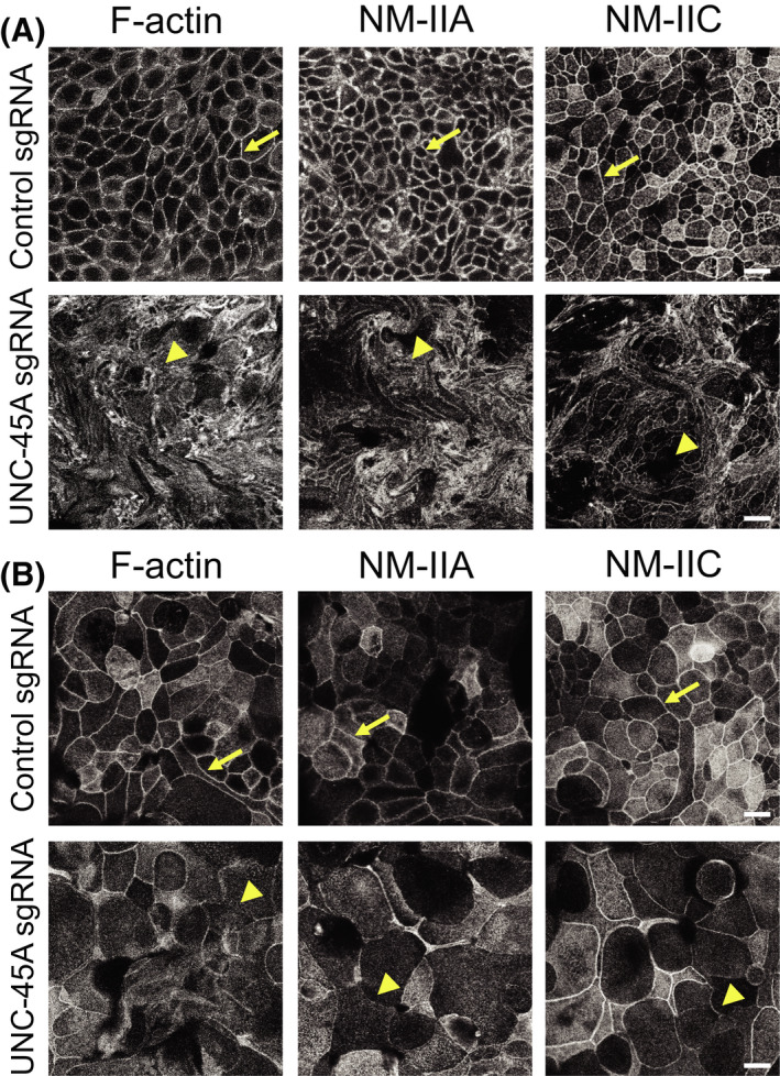
FIGURE 3.

Loss of UNC‐45A expression impairs organization of the perijunctional actomyosin belt in intestinal epithelial cell monolayers. Confocal microscopy images of control and UNC‐45A‐depleted HT‐29cf8 (A) and SK‐CO15 (B) cell monolayers fluorescently labeled for F‐actin and major epithelial myosin II motors, NM‐IIA and NM‐IIC. Arrows indicate perijunctional actomyosin structures in control IEC. Arrowheads show disruption of perijunctional actomyosin bundles in UNC‐45A‐depleted IEC. Scale bars, 20 µm. Representative of at least three independent experiments with multiple images taken per slide
