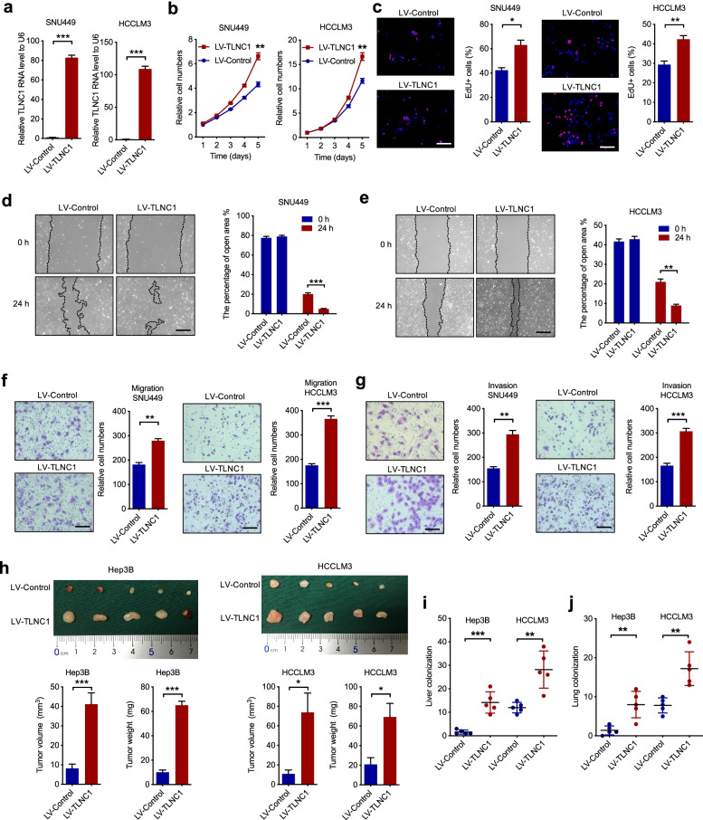
Fig. 2.
TLNC1 promotes tumor growth and metastasis of hepatoma cells. a qPCR quantification of the expression levels of TLNC1 in SNU449 and HCCLM3 cells stably transfected with lentivirus containing control or TLNC1 vectors. b Cell viability of indicated SNU449 and HCCLM3 cells was measured by CCK-8 assay. c Cell proliferation of indicated SNU449 and HCCLM3 cells was examined by EdU assay. d-e Cell migration of indicated SNU449 and HCCLM3 cells was measured by wound healing assay. f-g Cell migration and invasion of indicated SNU449 and HCCLM3 cells was measured by transwell migration and matrigel invasion assays. The data are the means ± SEM and are representative of three independent experiments. h Effects of TLNC1 overexpression in indicated Hep3B and HCCLM3 cells on subcutaneous tumor growth. Tumor volumes and tumor weights were measured after the mice were killed (n = 5). i The number of tumor nodules formed in the livers of orthotopic-implantation models inoculated with indicated hepatoma cells was indicated in the bar graph (n = 5). j The number of metastatic foci formed in the lungs of lung metastasis models inoculated with indicated hepatoma cells was indicated in the bar graph (n = 5). *p < 0.05, **p < 0.01, ***p < 0.001

