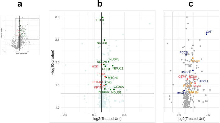
Figure 1.
Volcano plots of quantitative proteomic analysis of hearts of control and 2DG treated mice. (a) In miniature, fold-change and significance of all 1237 proteins identified in hearts of 2DG-treated mice versus controls. (b) Magnified region showing the significantly altered individual proteins related to the respiratory chain (green) and glycolysis (HXK1, PFKAM, PGK1, TPIS and KPYM) (red) (see Table S4 for the complete list and individual scores). (c) Magnified region of the volcano plot showing the significantly altered proteins related to mitochondrial branched-chain amino acid catabolism BCAT2, HMGCL, HIBCH, and PCCB, in blue, and OAT (which is involved in amino acid inter-conversion, as well as synthesis); 3 proteins in orange related to mtDNA expression (RRF2M, RM44, FAKD4), whereas three proteins of fatty acid metabolism, HCDH, MCAT and CBR4, are marked in red. Other components of the mitochondrial proteome significantly altered by the 2DG treatment (not highlighted) were two proteins related to mitochondrial morphology and division MTFP1, ARMC1, the mitochondrial transporter protein ABCB10; TOM70 a mitochondrial protein import factor; CLYBL (putative malate synthase); CDS2 (cardiolipin synthesis); GSHR and TRXR2 (redox homeostasis) and OXNAD1 (unknown function).

