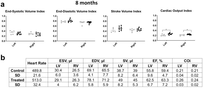
Figure 3.
Heart function is similar in control and 2-DG treated mice. (a) Heart capacity was measured by MRI. Right and left ventricle End-Systolic and End-Diastolic Volume index, Stroke Volume index and Cardiac Output index, adjusted for body surface area (BSA = 20 × body weight (g) ^ 0.42). There was no significant difference in the Ejection Fraction (EF) (amount, or percentage, of blood that is ejected from the ventricles with each contraction (EF = (SV/EDV) × 100)), or the volume remaining after contraction (End-Systolic Volume (ESV) or systole). There was a small increase in the cardiac output index in the 2DG treated animals (n = 8) versus controls (n = 6) which was significant, p = 0.03 for the right ventricle and p = 0.02 for the left. (b) Selected parameters measured to assess cardiac function. Heart rate (beats per minute), End-Systolic volume (ESV), End-Diastolic volume (EDV): volume of blood in a ventricle at the end of diastole, just before systole starts, Stroke volume (SV): volume of blood pumped from the left ventricle per beat (SV = EDV-ESV), Ejection Fraction (EF) (amount, or percentage, of blood that is ejected from the ventricles with each contraction (EF = (SV/EDV) × 100)), and Cardiac Output index (COi). The last was derived from Cardiac Output (CO): amount of blood pumped by the heart per minute (CO = (Heart rate x SV)/1000) and the Cardiac Index (CI), which relates the cardiac output (CO) from left ventricle in one minute to body surface area (CI = (CO/BSA) × 10,000)). There were no significant differences in the EF, ESV, CO or CI.

