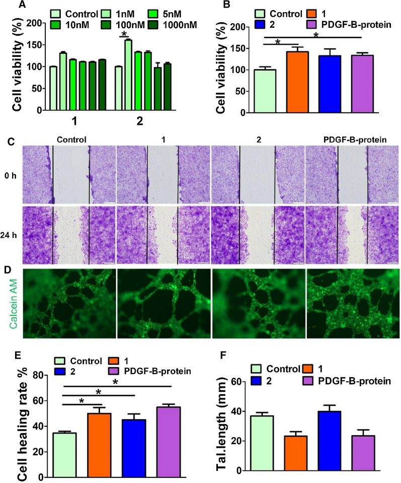
Fig. 3.
Bioactivity of PDGF mimicking peptides. A Cell viability of HUVEC cells incubated with 1 and 2 for 24 h. B Cell viability of HUVEC cells in serum-free culture medium containing 1 nM of 1, 2 or PDGF-B protein for 24 h. Data presented as the mean ± SEM, n = 3 samples per group. C Representative images of HUVECs after treated with 1, 2 or PDGF-B protein (1 nM) for 24 h. The edge of bilateral cell migration was marked with a black line. Image was taken at 10× magnification, scale bar = 100 μm. D Microvessel formation assay, HUVECs incubated with 1, 2 or PDGF-B protein (1 nM) for 6 h, then stained with Calcein AM. Scale bar = 100 μm. E Quantification of HUVECs cell healing rate, which was determined by image J. F The counts of branching interval quantification of HUVECs was determined by image J. Angiogenesis.*p < 0.05 V.S. control group

