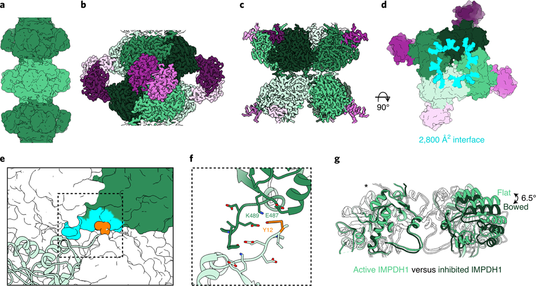
Fig. 3 |. Inhibited IMPDH1 assembles with an alternative filament architecture (gtP/AtP/IMP bound).
a, Low-pass filtered cryo-EM reconstruction colored by octamer. b, Octamer-centered single-particle reconstruction at 2.6 Å of the filament, with catalytic domains in different shades of green and regulatory domains in shades of pink. c, Interface-centered single-particle reconstruction of the filament at 2.6 Å. d, View of the top of an octamer from inside the filament. Surface representation of the atomic model at the assembly interface, with buried residues in cyan. e, Surface representation of filament interface with one monomer in ribbon (light green). Tyr12 is shown in orange spheres. The monomer it contacts across the octamer interface is green with residues forming the interface in aqua. f, Close-up ribbon view of the interface where Tyr12 in orange contacts Glu487 and Lys489 in the opposing monomer. g, Comparison of the catalytic tetramers of active IMPDH1 (light green) to mostly inhibited IMPDH1 (dark green), show that the GTP-bound structure is in the bowed conformation. Aligned on monomers with an asterisk.

