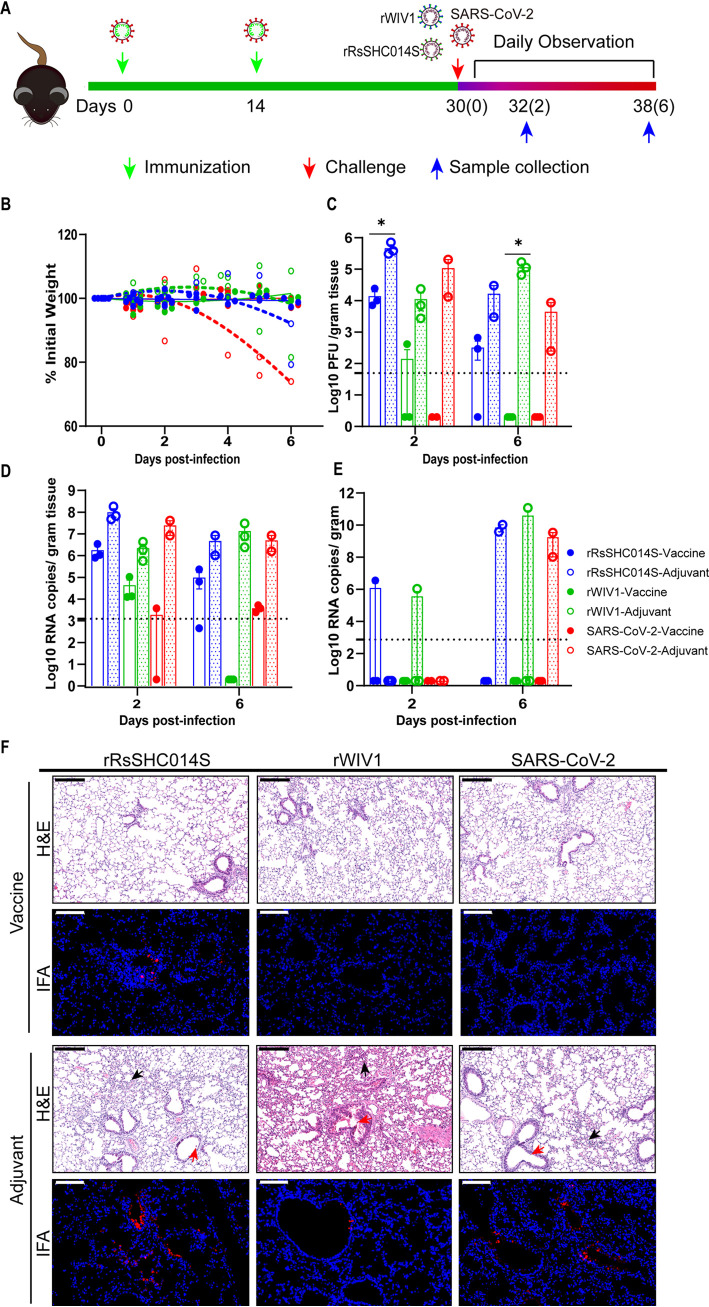
FIG 1.
SARS-CoV-2 IAV partially protects mice from bat SARSr-CoV infection. HFH4-hACE2 mice were intraperitoneally immunized with 5 μg of SARS-CoV-2 IAV and 0.5 mg of aluminum hydroxide (vaccine group) or 0.5 mg of aluminum hydroxide with PBS (adjuvant group) following a D0/D14 immunization program. At 30 days after the initial injection, the mice were infected with 105 PFU of the indicated virus. (A) Experimental scheme. (B) Mouse body weight was monitored for up to 6 dpi. Dotted lines represent the fitted curves for each color-indicated group. Error bars indicate standard errors. (C) Lung viral loads as detected by plaque assays. (D) Lung viral loads as detected by qRT-PCR. (E) Brain viral loads as detected by qRT-PCR. The dotted line indicates the limitation of qRT-PCR detection. (F) Lung pathological changes and viral antigen staining at 2 dpi. Bronchiolar epithelial sloughing (red arrows) and few infiltrations (black arrows) were observed in all mice in the adjuvant groups. Especially, alveolar wall thickening was observed in the rWIV1 adjuvant group. Few pathological changes in the lungs were observed in the vaccinated groups. Viral antigen was detected in all adjuvant groups, whereas no distinct signal was detected in vaccinated mice challenged with SARS-CoV-2 and rWIV1. A weak antigen signal was detected in the lungs of vaccinated mice challenged with rRsSHC014S. Images were acquired using a Pannoramic MIDI system. Black scale bar, 200 μm; white scale bar, 100 μm. Error bars indicate standard errors. Statistical significance was assessed using the Mann-Whitney test (*, P < 0.05).

