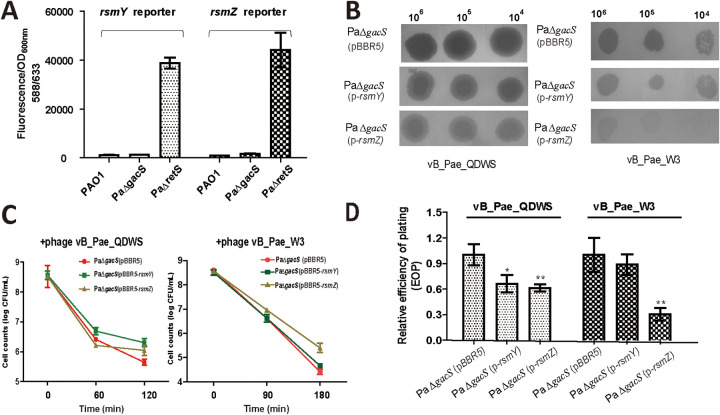
FIG 3.
RsmY/Z levels were closely associated with phage resistance. (A) P. aeruginosa PAO1, PaΔgacS, and PaΔretS cells containing pBBR-PrsmY-mkate or pBBR-PrsmZ-mkate grew in LB medium until an OD600 of 1 and were collected for fluorescence assays. (B) A total of 3 μL of serial dilutions of vB_Pae_QDWS and vB_Pae_W3 were spotted onto PAO1 (pBBR5), PaΔgacS (p-rsmY), and PaΔgacs (pBBR5-rsmZ) for lytic activity assays. (C) Cell counts of PaΔgacS (pBBR5)(●, red mark), PaΔgacS (p-rsmY)(■, green mark), and PaΔgacS (p-rsmZ)(▲, yellow mark) strains infected with phage vB_Pae_QDWS or vB_Pae_W3 at an MOI of 1 were detected at different time points. (D) Relative efficiency of plating (EOP) of phage vB_Pae_QDWS and vB_Pae_W3 on P. aeruginosa strains. The values were the averages of three measures with standard deviation. Symbol * indicates the sample is different (0.01 < P < 0.05) from the control PaΔgacS (pBBR5), and symbol ** indicates the sample is significantly different (P < 0.01) from PaΔgacS (pBBR5) (Student’s paired t test).

