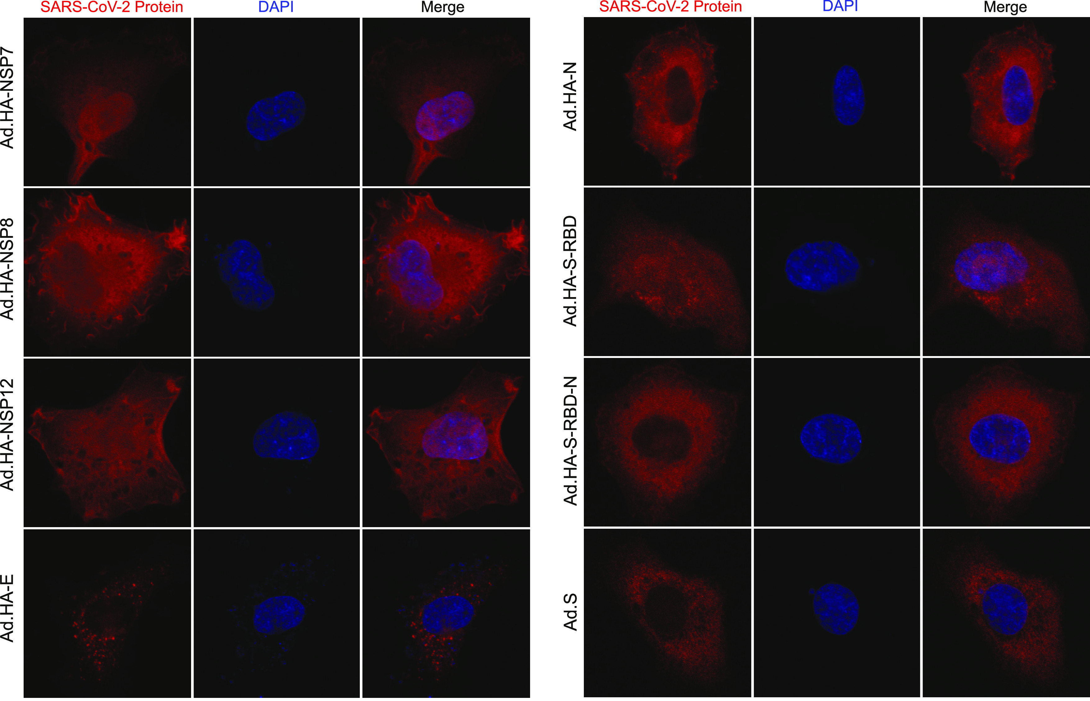
FIG 5.

Subcellular localization of SARS-CoV-2 protein expressed from rHAdV vectors. HT1080 cells were infected with the indicated viruses at an MOI of 1,000 vp/cell. Then, 24 h after infection, cells were fixed and stained either for HA using the rat monoclonal 3F10 or for S using 2B3E5. DAPI was used as a nuclear counterstain, and representative images for each protein are shown. Images were acquired on a Zeiss LSM700 laser confocal microscope using a 63× lens objective.
