FIG 2.
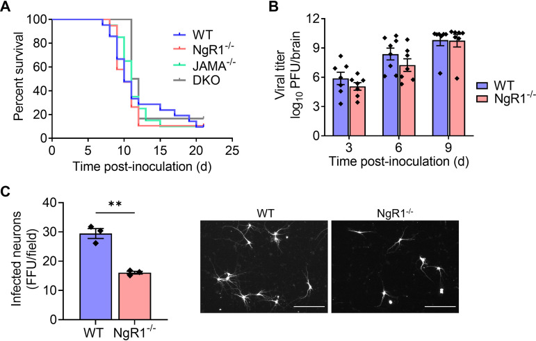
NgR1 is not required for reovirus neuropathogenesis following intracranial inoculation. (A and B) Newborn WT, JAM-A−/−, NgR1−/−, and DKO mice were inoculated intracranially with 25 PFU of reovirus T3SA−. (A) Mice were monitored for disease signs and survival for 21 days and euthanized when moribund. n = 6 to 21 mice per group. Differences in survival relative to WT mice were not significant, as determined by log-rank test. (B) At 3, 6, and 9 days after inoculation, mice were euthanized, brains were removed and hemisected, and viral titers in homogenates of the inoculated half of the brain were determined by plaque assay. Results are expressed as mean viral titers. Error bars indicate SEM. n = 8 to 11 mice per group for each time point. Differences in titer between WT and NgR1−/− mice at each time point were not significant (P > 0.05), as determined by two-way ANOVA with Dunnett’s multiple-comparison test. (C) Cortical neurons isolated from E15.5 WT or NgR1−/− mice were adsorbed with T3SA+ virions at an MOI of 500 PFU/cell. Cells were fixed at 24 h postadsorption and stained using reovirus-specific antiserum. Results are expressed as the mean number of infected neurons, identified based on morphology, per field-of-view. Error bars indicate SEM for triplicate samples from one representative experiment of two conducted. **, P < 0.01 (t test). Representative micrographs on the right show infected neurons in an area approximately one-fourth of the field-of-view examined. Bars, 150 μm.

