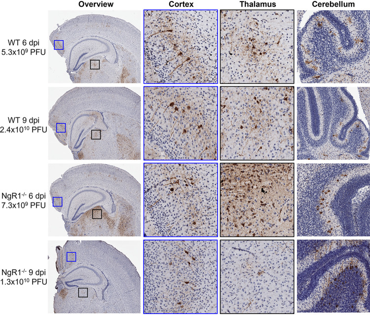
FIG 3.
Reovirus tropism in the brain is unaltered in the absence of NgR1. Newborn WT and NgR1−/− mice were inoculated intracranially in the right brain hemisphere with 25 PFU of reovirus T3SA−. Mice were euthanized 6 or 9 dpi, and brains were removed and hemisected. Left-brain hemispheres were fixed in formalin and embedded in paraffin. Coronal sections of the left-brain hemisphere were stained with reovirus-specific antiserum and hematoxylin. Representative sections show reovirus antigen in cortex, thalamus, and cerebellum. Enlarged images of areas boxed in the overview show reovirus infection of neurons in the cortex (blue boxes) and thalamus (black boxes). Viral titers from the paired right-brain hemispheres are reported adjacent to the micrographs.

