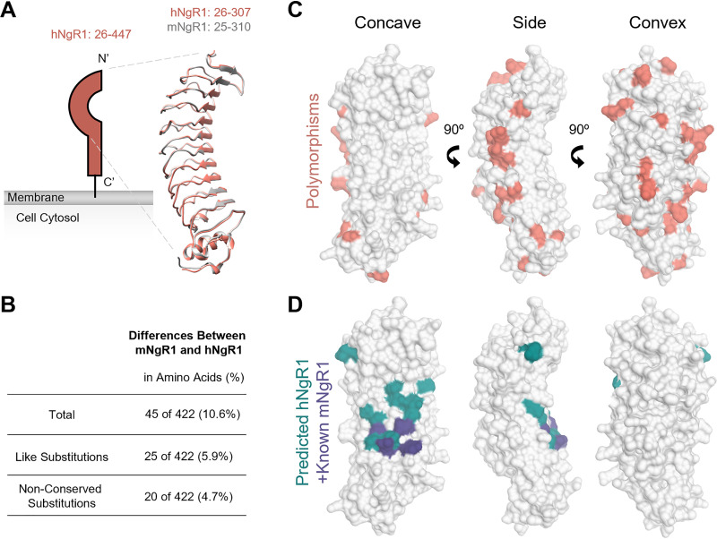
FIG 7.
Comparison of human and mouse NgR1 structures and binding surfaces. (A) Schematic of NgR1 alongside overlaid ribbon tracings of hNgR1 (PDB ID 1OZN) (52) and mNgR1 (PDB ID 5O0K) (53). Amino acids included in the model are indicated. N and C termini are labeled. (B) Comparison of hNgR1 and mNgR1 amino acid sequences. (C) Surface representations of hNgR1 amino acids 26 to 307 (PDB ID 1OZN), with mNgR1 polymorphisms shown in coral. (D) Surface representations of hNgR1, with residues identified by alanine mutagenesis predicted to be required for the binding of hNgR1 by neural ligands (39) shown in teal. Residues required for the binding of mNgR1 by BAI1 (40) are shown in purple. Structure representations were made using Chimera UCSF (54).

