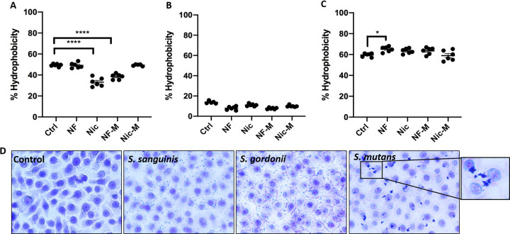
FIG 5.
S. mutans shows a higher hydrophobicity and coaggregation capacity than S. sanguinis and S. gordonii. Overnight cultures of S. sanguinis (A), S. gordonii (B), and S. mutans (C) were diluted 1:10 in e-cig-pretreated medium and grown for 24 h. Chloroform was added to each condition, and after incubation, the aqueous layer was collected and OD600 was measured. The data are means and SEM) (n = 3) for three biological replicates. Groups were compared to controls using one-way ANOVA (Dunnett’s correction). *, P < 0.05; ****, P < 0.0001. (D) Bright-field microscopy of crystal violet-stained 1-h cocultures (MOI, 10) of oral epithelial cells (OKF6) with S. sanguinis, S. gordonii, and S. mutans. Magnification, ×40.

