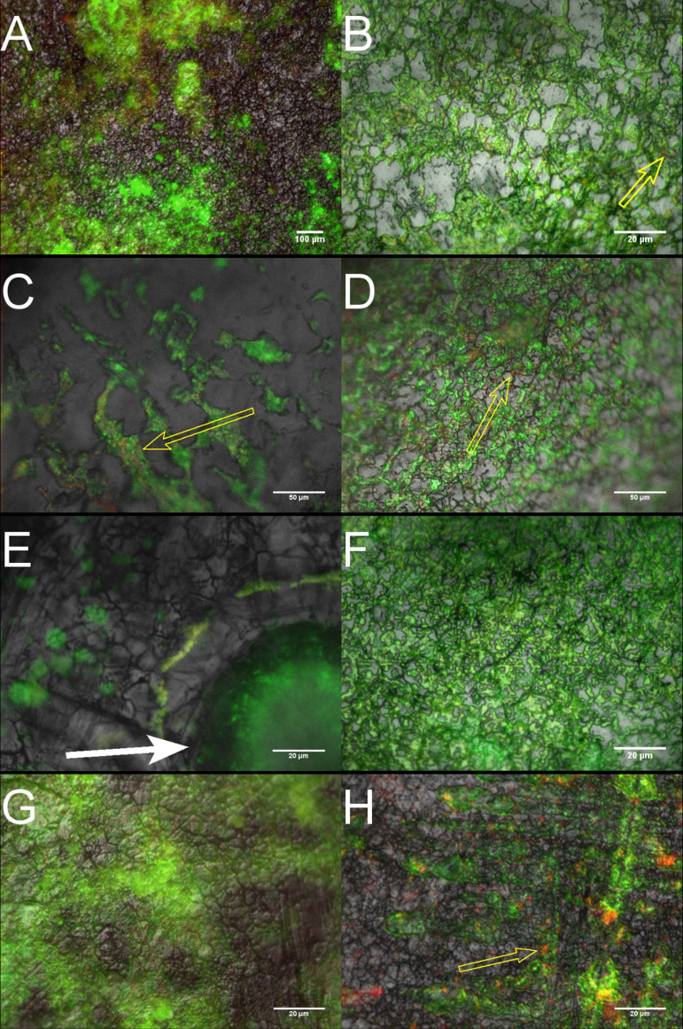
FIG 1.
Representative EDIC/EF images of biofilm formation by A. baumannii (A and B), S. aureus (C and D), E. faecalis (E and F), and P. aeruginosa (G and H) on stainless steel coupons. Left-hand side micrographs are cultures grown under AHS; right-hand side, cultures grown in nutrient broth. The micrographs demonstrate the distinct difference in microcolony distribution between the two medium types; most notably, the spatial arrangement of colony niches around the artificial sebum aggregates, indicated by the white solid arrows. Traces of nonviable bacterial cells can be seen within the biofilms of both media types, highlighted by yellow outlined arrows.

