FIG 3.
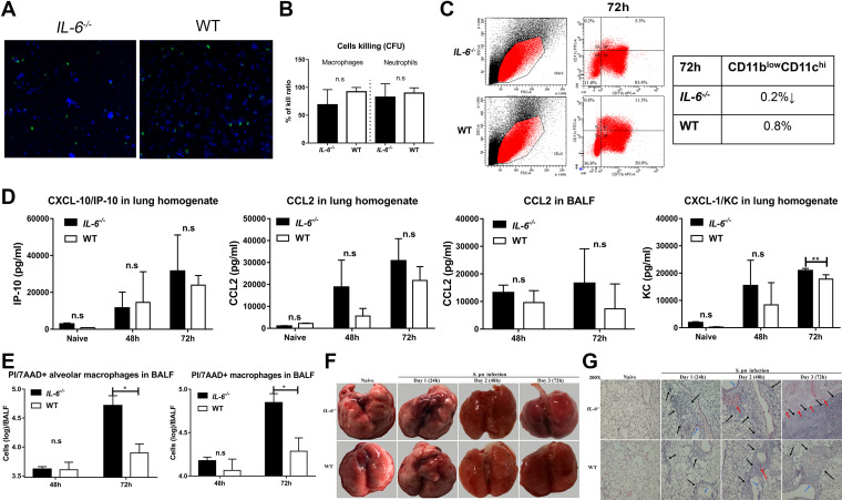
IL-6 is crucial for lung macrophage survival during pneumococcal pneumosepsis. (A) In vivo phagocytosis assays of mice (n = 3/group). (B) In vitro CFU-based S. pneumoniae killing analysis of peritoneal macrophages and neutrophils (n = 4 to 9/group). (C) Percentage of CD11blowCD11chi cells in BALF of mice during pneumococcal pneumosepsis (n = 3/group). (D) CXCL-10, CCL2, and CXCL-1 levels in lungs of mice at 48 and 72 hpi were measured by ELISA, (n = 4 to 6/group). (E) Propidium iodide/7-aminoactinomycin D (7-AAD+)-staining of alveolar and resident lung macrophages of IL-6−/− and WT mice at 48 and 72 hpi (n = 4 to 5/group). (F to G) Representative photomicrographs of lung tissue histopathology (n = 3/group) and H&E-stained tissues (n = 3/group) of IL-6−/− and WT mice from 1 to 3 dpi. Black arrow, inflammatory cell infiltration; blue arrow, epithelial cell shedding; red arrow, bleeding.

