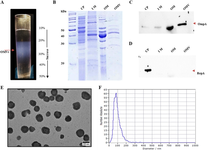
FIG 1.
Purification of outer membrane vesicles (OMVs) from Acinetobacter baumannii DS002. (A) OMV band on sucrose density gradient (10 to 50%). (B) SDS-PAGE (12%) showing the profiles of cytoplasmic (CP), inner membrane (IM), outer membrane (OM), and OMV proteins. Corresponding Western blots, developed by probing with either OmpA or RepA antibodies, are shown in panels C and D, respectively. Panel E shows transmission electron microscopy (TEM) images of pure OMVs, showing their size distribution. Size distribution of OMVs as measured by particle matrix analyser (ZetaView) is shown in panel F.

