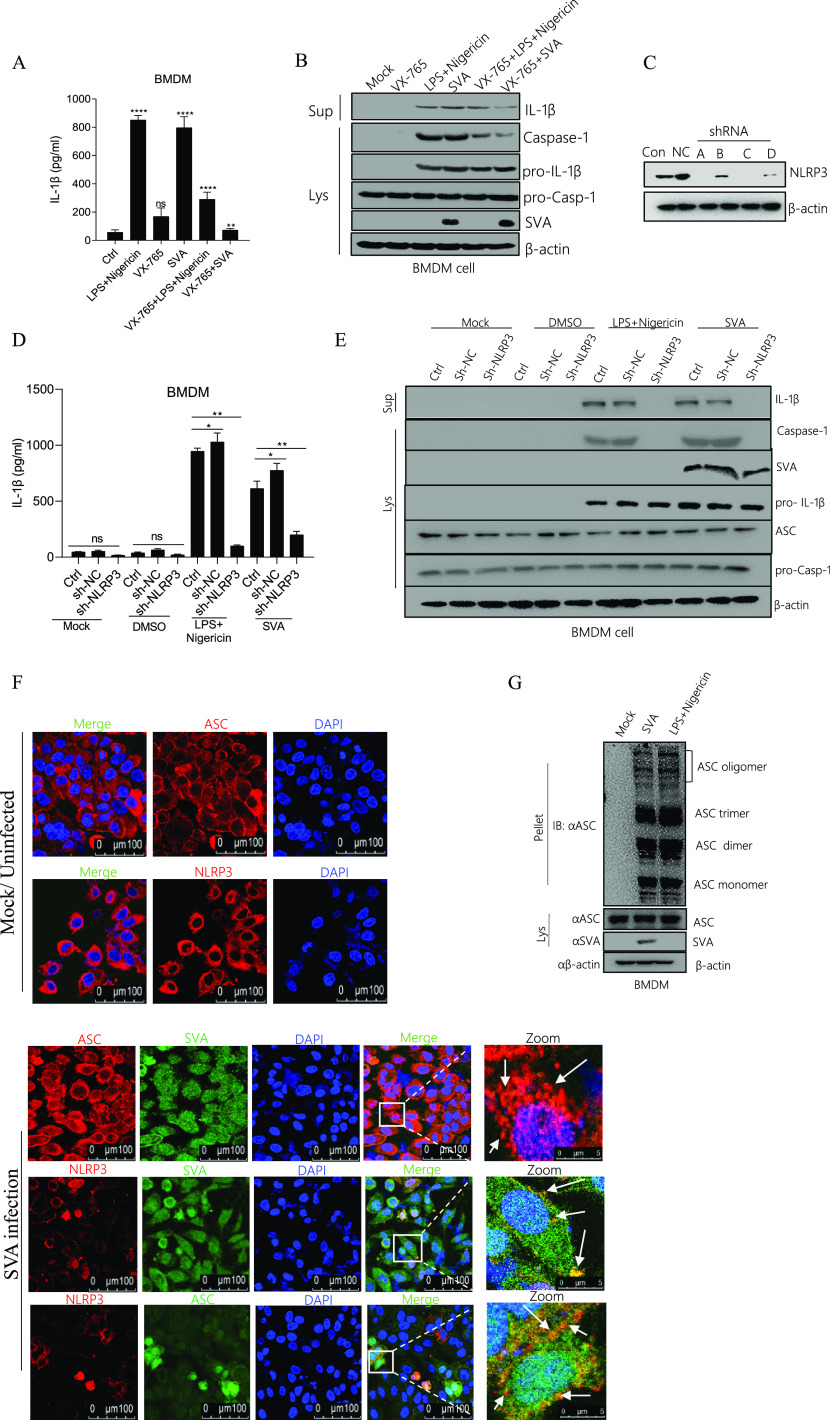
FIG 2.
SVA activates the NLRP3 inflammasome to induce IL-1β secretion. (A, B) Porcine BMDM cells were treated with 2 μM Nigericin (NLRP3 stimulator) for 2 h pre-primed with LPS (60 ng/mL) for 8 h or 10uM VX-765 (casp-1 inhibitor) for 1 h or infected with SVA at MOI = 4 for 16 h or 10uM VX-765 together with 2 μM Nigericin and LPS (60 ng/mL) or 10uM VX-765 together with SVA infection. The IL-1β levels in the medium were determined by ELISA (A). IL-1β (17 kDa) expression in supernatants or pro-IL-1β (31 kDa), Caspase-1 (P20), and pro-casp-1 (45 kDa) expression in lysates were detected by Western blotting (B). (C) Porcine BMDM cells were infected with lentivirus-shRNAs targeting NLRP3 or sh-NC (negative control), and after 48 h, NLRP3 protein expression was detected by Western blotting. (D, E) Porcine BMDM cells were infected with lentivirus-shRNA targeting NLRP3 (shNLRP3-A) and treated with DMSO or 2 μM Nigericin for 2 h pre-primed with LPS (60 ng/mL) for 8 h or infected with SVA at MOI = 4 for 16 h. The IL-1β levels in the medium were determined by ELISA (D). IL-1β (17 kDa) expression in supernatants or pro-IL-1β (31 kDa), Caspase-1 (P20), ASC (22kDA), SVA, and pro-casp-1 (45 kDa) expression in lysates were detected by Western blotting (E). (F) Porcine BMDM cells were infected with SVA at MOI = 4 for 16 h. NLRP3, ASC, and SVA subcellular localizations were assayed by confocal microscopy. The scale bar was 100 μm, and the zoom section was 5 μm. (G) Porcine BMDM cells were infected with SVA at MOI = 4 for 16 h or treated with 2 μM Nigericin for 2 h pre-primed with LPS (60 ng/ml0 for 8 h). ASC oligomerization with ASC primary antibody was detected by Western blotting. For Western blot, the antibody dilution ratio was 1:1,000. The data shown are the mean±s.e.m. *, P < 0.05; **, P < 0.01; ***, P < 0.001; ****, P < 0.0001 versus mock (one-way ANOVA with Tukey's post hoc test). All experiments were repeated three times with similar results. Data were representative of the three independent experiments.

