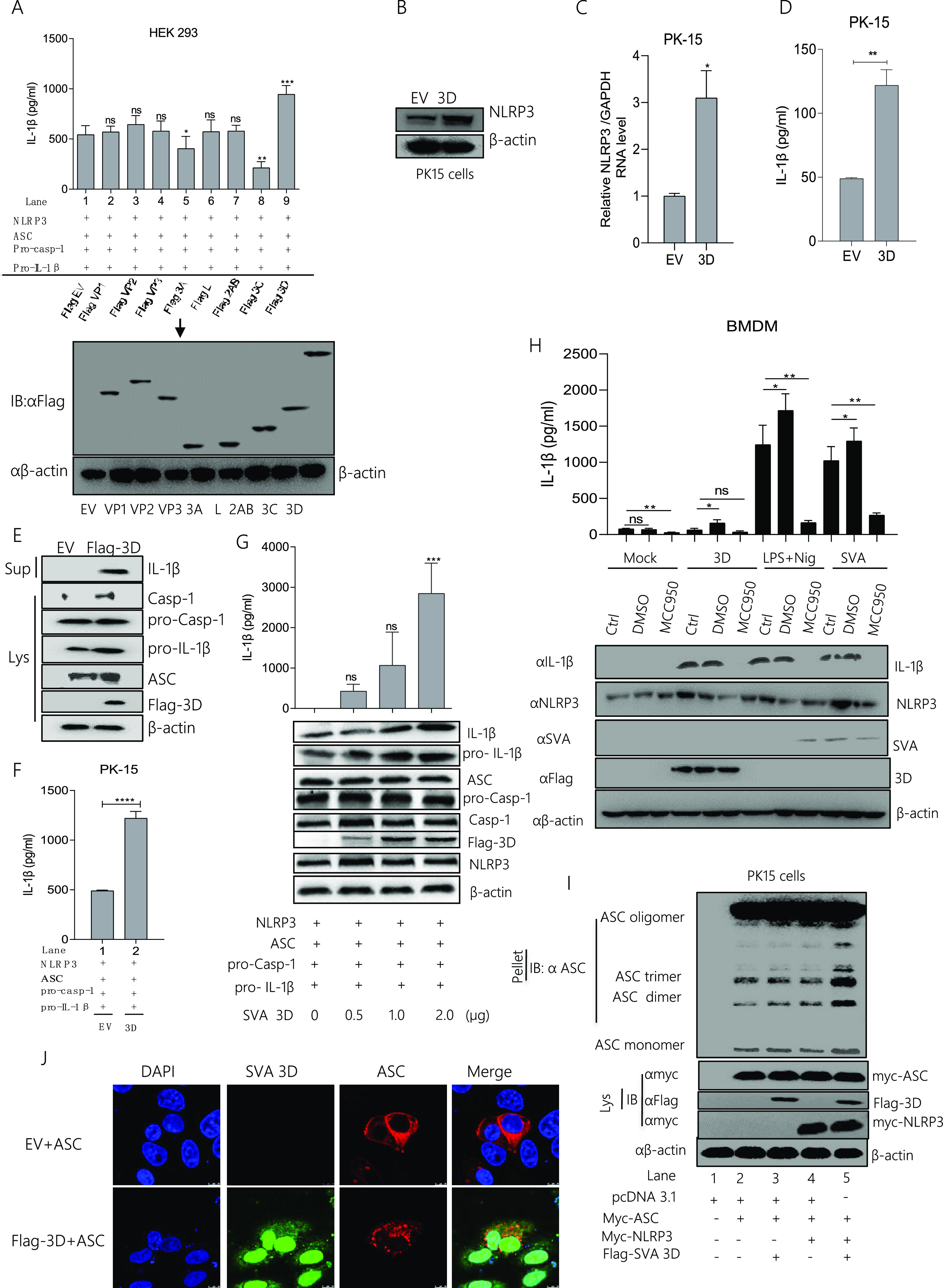
FIG 4.

SVA polymerase 3D facilitates NLRP3-mediated IL-1β production. (A) HEK293 cells were co-transfected with 2 μg of plasmids encoding NLRP3, ASC, pro-casp-1, and pro-IL-1β. The cells were then transfected with 2 μg of pcDNA 3.1 as an empty vector, Flag VP1, Flag VP2, Flag VP3, Flag 3A, Flag L, Flag 2AB, Flag 3C, and Flag 3D separately with the above-indicated plasmids. IL-1β levels from the medium were detected by ELISA, and all the SVA plasmids (Flag VP1, Flag VP2, Flag VP3, Flag 3A, Flag L, Flag 2AB, Flag 3C, and Flag 3D) expressions were detected by Western blotting (A). (B to E) PK-15 cells were transfected with 2 μg Flag-3D or 2 μg empty vector pcDNA 3.1. Cell lysates were subjected to SDS-PAGE, NLRP3 (110 kDa) was detected by Western blotting (B), and NLRP3 mRNA levels were detected qPCR (C), IL-1β levels from medium were detected by ELISA (D). IL-1β (17 kDa) expression in supernatants or Caspase-1 (P20), pro-IL-1β (31 kDa), ASC (22kDA), Flag 3D (51 kDa), and pro-casp-1 (45 kDa) expression in lysates were detected by Western blotting (E). (F) PK-15 cells were co-transfected with 2 μg plasmids encoding NLRP3, ASC, pro-casp-1, and pro-IL-1β and then transfected with 2 μg Flag 3D or 2 μg empty vector pcDNA 3.1. IL-1β levels from the medium were detected by ELISA. (G) PK-15 cells were co-transfected with NLRP3, ASC, pro-casp-1, and pro-IL-1β and transfected with plasmids expressing the Flag 3D of SVA in a dose-dependent manner (0, 0.5, 1.0, 2.0 μg). IL-1β levels were detected by ELISA and IL-1β (17 kDa) expression in supernatants or pro-IL-1β (31 kDa), ASC (22 kDa), Flag 3D (51 kDa), mature Caspase-1 (20 kDa), NLRP3 (110 kDa), and pro-casp-1 (45 kDa) expression in lysates were detected by Western blotting. (H) The BMDM cells were transfected with 2 μg of 3D, postinfected with SVA or post-stimulated with LPS (60 ng/mL) and Nigericin (2 μM), and the cells were untreated or treated with DMSO or 5 μM MCC950 (NLRP3 activation inhibitor) for 16 h. Cell lysates were subjected to Western blotting, IL-1β (17 kDa) expression in supernatants or NLRP3 (110 kDa), SVA, and Flag 3D (51 kDa) expression in lysates were detected by Western blotting and also IL-1β levels from medium were detected by ELISA. (I) PK-15 cells were co-expressed with 2 μg of pcDNA 3.1, myc-ASC, myc-NLRP3, or Flag 3D. Cell lysates and pellets were subjected to ASC oligomerization detection. (J) PK-15 cells were transfected with EV+ ASC, Flag-3D+ ASC. Subcellular localization was observed by confocal microscopy, and the scale bar is 10 μm. After transfection, samples were harvested for 24 h. For Western blot, the antibody dilution ratio was 1:1,000. The number of replicates is three. Data shown are mean±s.e.m.; *, P < 0.05; **, P < 0.01; ***, P < 0.001; ****, P < 0.0001 vs EV; ns, no significance (one-way ANOVA with Tukey’s post hoc test).
