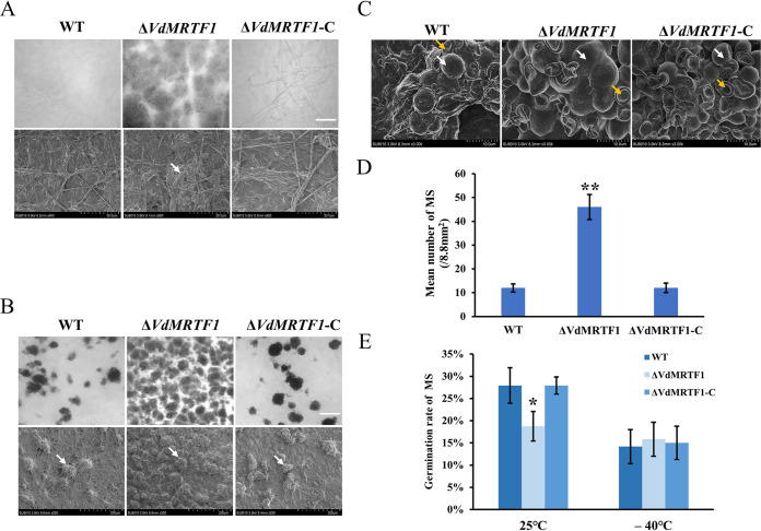
FIG 2.
Microsclerotia formation, germination, and morphology of wild-type, ΔVdMRTF1, and ΔVdMRTF1-complemented strains of Verticillium dahliae. (A) Microsclerotium formation of the wild-type, ΔVdMRTF1, and complemented (ΔVdMRTF1-C) strains were captured by biological microscope (above) and scanning electron microscopy(below) on BM at 25°C for 4 days. White arrow points to a microsclerotium. Bar = 100 μm. (B) Microsclerotium formation of the wild-type, ΔVdMRTF1, and ΔVdMRTF1-C strains were captured by biological microscope (above) and scanning electron microscopy(below) on BM at 25°C for 7 days. White arrow points to a microsclerotium. Bar = 100 μm. (C) The morphology of 14-day-old microsclerotia incubation on BM by scanning electron microscopy. White arrow points to plump microsclerotia, and the orange arrow points to widened microsclerotia. (D) The bar chart showed the mean number of microsclerotium (MS) of all strains on BM at 25°C for 7 days. Error bars represent the standard deviations of three replicates. Asterisks indicate significant differences (**P < 0.01). (E) The bar chart showed the germination rate of microsclerotium (MS) of the wild-type, ΔVdMRTF1, and ΔVdMRTF1-C strains in different temperature. Error bars represent the standard deviations based on three independent replicates. Asterisks indicate significant differences (*P < 0.05).

