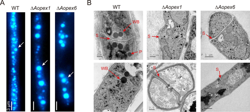
FIG 5.
Observation of autophagosomes and ultrastructure of peroxisomes and Woronin bodies in the wild-type (WT) and mutant strains. (A) The autophagosomes of WT strain, ΔAopex1, and ΔAopex6 mutant strains were stained with MDC. Bar: 3 μm. (B) The WT and mutant strains were observed by transmission electron microscopy. WB: Woronin body; P: peroxisome; S: septum.

