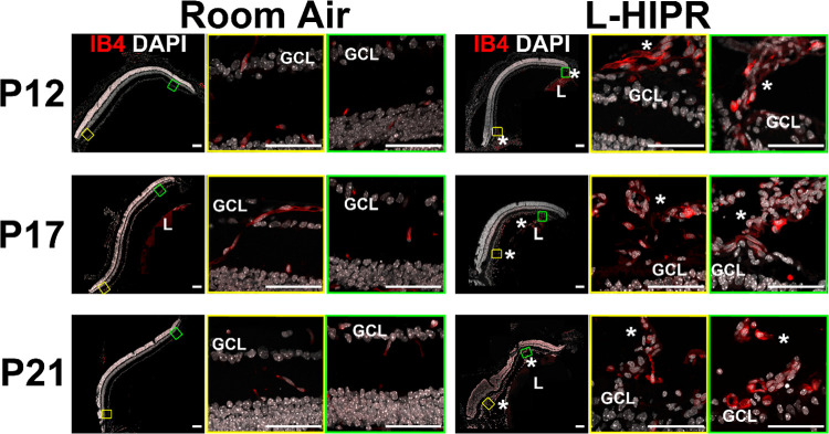
Fig 4. The hyaloidal vessels persist and rescue the retinal vasculature in L-HIPR.
Retinal cross sections in room air and L-HIPR groups (n = 3) were stained with Isolectin B4 (IB4; red), and DAPI (white) at P12, P17 and P21. Central (yellow box) and peripheral (green box) zoomed retinal cross-sections are presented. The hyaloidal vessels were absent, with normal retinal vascular stratification by P12 in the room air eyes. In L-HIPR, the hyaloidal vessels persisted well into P21 and started to invade the peripheral retina at P12 (Asterisks). The later timepoints further illustrate how the hyaloidal vessels invade the mid-retina at P17 and P21. Scale bar 50 μm, GCL: ganglion cell, *: hyaloid, L: lens.

