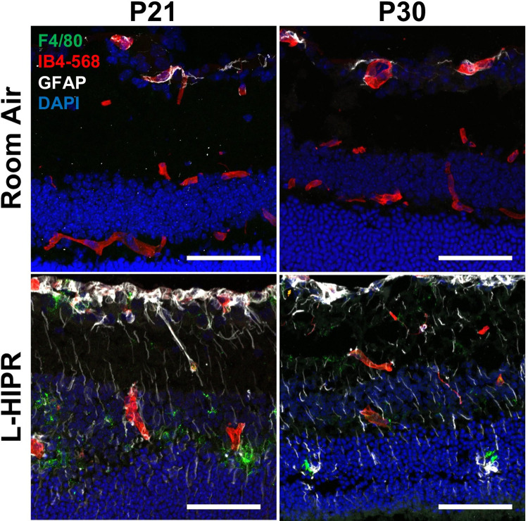
Fig 5. The L-HIPR retina is inflamed.
(A) Immunolabeled cryosections at P21 and P30 of room air and L-HIPR tissue. Macrophages were labeled with anti-F4/80 (green), blood vessels with IB4 (red), astrocytes/activated Muller cells with GFAP (white), and nuclei with DAPI (blue). The room air tissues show GFAP+ astrocytes in the ganglion cell layer, however, the Muller cells become GFAP+ indicating an activated state in L-HIPR at both P21 and P30. F4/80+ macrophages can be seen in the inner and outer nuclear layers in the L-HIPR sample. Scale bars equal 50 μm.

