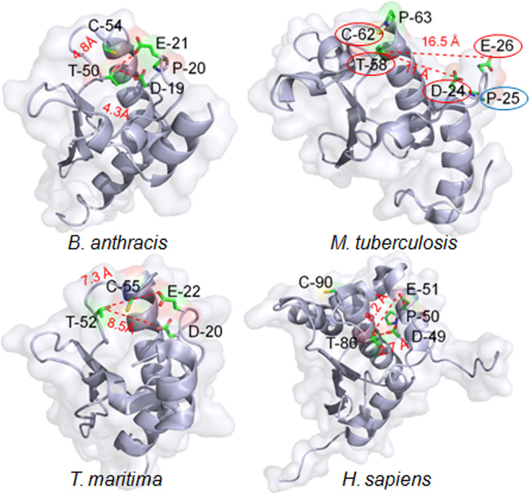
Fig 1. Three-dimensional structure of Rv1466 (SufT).
Mtb SufT like other DUF59 proteins contains a small surface of conserved homology. Ribbon diagrams are shown for the Bacillus anthracis YitW (PDB ID: 3LNO); Mtb SufT (PDB ID: 5IRD); Thermotoga maritima TM0487 (PDB ID: 1UWD); and Homo sapiens FAM96a (PDB ID: 2M5H). The side chains of the conserved D, E, T, and C (red encircled) residues are highlighted. Oxygen, sulfur, and nitrogen atoms are represented with red, yellow, and blue color, respectively. Distance in Angstrom (Å) is represented between the side-chain hydroxyl of the T, D, and E residues. A proline (P25 in Mtb) present in the motif as DPE (blue encircled) and the consecutive proline (P63 in Mtb) next to the C residue are represented in the Mtb SufT structure.

