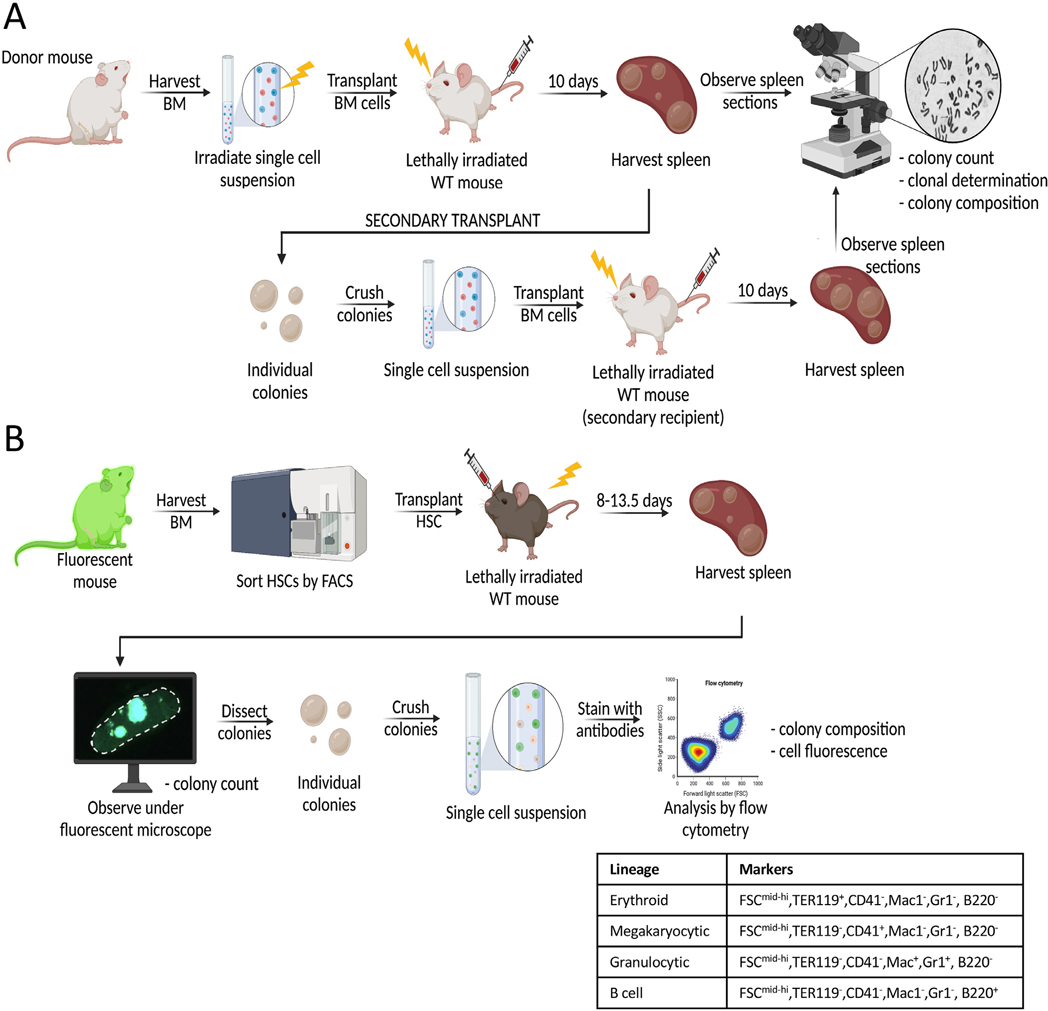
Figure 1.
Schematic of CFU-S assays. (A) In the original CFU-S assay developed by Till and McCulloch in 1961 [2], bone marrow cells were harvested from donor mice and irradiated prior to intravenous injection into irradiated recipient mice. Ten days post transplant, spleens were counted, harvested, and sectioned for histologic analysis. For serial transplantations to determine self-renewal of CFU-S cells within spleen colonies [15], individual colonies were dissected and transplanted into secondary recipients as single-cell suspensions. Spleen colonies formed after secondary transplantation were analyzed similarly to those from primary transplantations [15]. (B) In the updated CFU-S assay with high throughput, quantitative analysis, HSCs (or other hematopoietic progenitors) were FACS-purified and transplanted into irradiated recipient mice. At 13.5 days post transplan, individual spleen colonies were counted, harvested, and dissected under a fluorescent microscope [22]. Single-cell suspensions from each colony were then analyzed by flow cytometry to assess colony composition and cell fluorescence. Four lineages (erythroid, megakaryocytic, granulocytic, and B cell) were identified using the markers listed in the table [22,41]. BM=bone marrow; CFU-S=spleen colony-forming unit; HSCs=hematopoietic stem cells.

