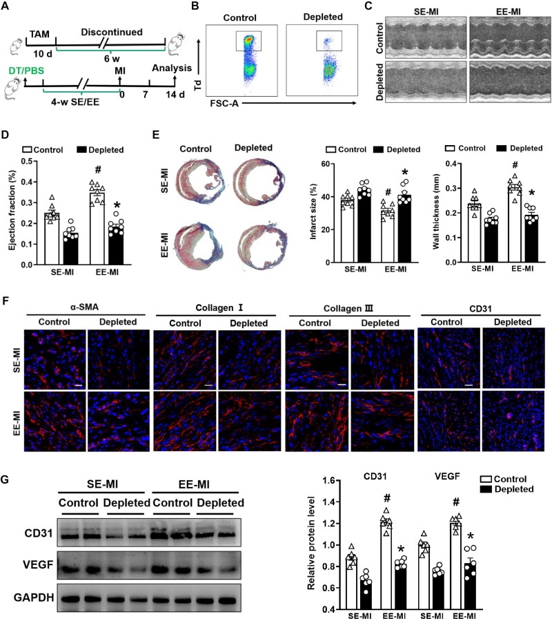
Fig. 6. Depletion of cardiac resident macrophages impairs the EE-mediated cardioprotective effect.
(A) Timeline diagram of the experimental procedures. (B) Representative flow cytometric plots of Td+ resident macrophages following the administration of DT to Cx3cr1CreER×R26Td/DTR and Cx3cr1CreER×R26Td/+ mice. (C and D) Echocardiographic analysis of EF value at day 14 after MI after SE and EE housing (n = 8). #P < 0.05 versus SE-MI. *P < 0.05 versus control. (E) Representative Masson’s staining of cardiac tissue obtained from depleted and control mice 14 days after MI. Quantitative analysis of infarct size and wall thickness (n = 8). #P < 0.05 versus SE-MI. *P < 0.05 versus control. (F) Immunostaining analyses of α-SMA, collagen I, collagen III, and CD31 on infarct sections collected at 14 days after MI. Scale bars, 20 μm (for α-SMA, collagen I, and collagen III) and 50 μm (for CD31). (G) Western blot analyses of VEGF and CD31 expression in the infarct tissues at day 14 after MI (n = 6). #P < 0.05 versus SE-MI. *P < 0.05 versus control. Data are expressed as means ± SEM. Data were analyzed using Student’s t test.

