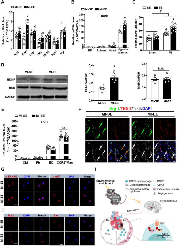
Fig. 7. EE promotes CCR2−MHCIIlow macrophage survival via the BDNF-TrkB axis.
(A) mRNA expression of agouti-related peptide (Agrp), Bdnf, leptin receptor (Lepr), neuropeptide Y (Npy), neurotrophic receptor tyrosine kinase 2 (Ntrk2), serum/glucocorticoid-regulated kinase1 (Sgk1), and nerve growth factor inducible (Vgf) in hypothalamus obtained from SE and EE mice at day 7 after MI (n = 6). *P < 0.05 versus SE. (B) mRNA expression of BDNF in bone marrow (BM), spleen, heart, and hypothalamus collected from SE and EE mice at day 7 after MI (n = 6). *P < 0.05 versus SE. (C) The BDNF content in plasma of SE- and EE-housed mice at day 14 after MI or sham operation. *P < 0.05 versus SE and #P < 0.05 versus sham. (D) Western blot analyses of BDNF and TrkB expression in the infarct tissues at day 14 after MI (n = 6). *P < 0.05 versus MI-SE. (E) TrkB mRNA expression in cardiomyocytes (CMs), fibroblasts (Fb), endothelial cells (ECs), and CCR2−MHCIIlow macrophages sorted from the hearts at day 7 after MI (n = 6). *P < 0.05 versus MI-SE. (F) Immunostaining analyses of Arg-1, cardiac troponin I (TNNI3), and TrkB on infarct sections collected at 14 days after MI. Scale bars, 20 μm. (G) Immunostaining of p-ERK1/2 and p-AKT in CCR2−MHCIIlow macrophages sorted from infarcted hearts. Scale bars, 50 μm. (H) Immunostaining of Bcl-2 and Bax in CCR2−MHCIIlow macrophages sorted from the infarcted hearts. Scale bars, 50 μm. (I) Mechanism of EE-induced cardioprotective effect in mice after MI.Data are expressed as means ± SEM. Data in (C) were analyzed using two-way ANOVA followed by Bonferroni post hoc analysis. Data in (A), (B), (D), and (E) were analyzed using Student’s t test.

