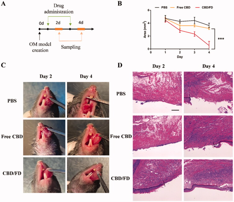
Figure 5.
The therapeutic effect of CBD/FD nanomicelles on tongue ulcers after intravenous administration. (A) Drug administration and sampling. (B) Change in the ulcer area over time (n = 5). Data are shown as mean ± SD. (C) Tongue ulcer images at 2 and 4 days post-administration. (D) Hematoxylin and eosin images of tongue ulcers. Solid lines indicate the ulcer boundary, and dotted lines indicate the epithelial-stromal boundary. Scale bar, 100 μm.

