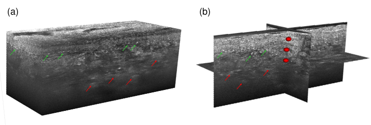
Fig. 6.
(a) 3D LC-OCT image and (b) orthogonal views of a red-colored tattoo biopsy with multiple POIs materialized by red ellipsoids. Diffuse bright areas in the dermis can be observed in the LC-OCT image, depicted by red arrows. Hyper-reflective structures of about 10-40 in size can be noticed near the dermal-epidermal junction, pointed out by green arrows.

