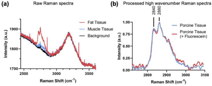
Fig. 4.
(a) Raw Raman spectra of porcine fat/muscle tissue acquired through the coherent fiber bundle during CLE imaging. These have no background subtraction or smoothing. Also shown is the background signal of the system (b) Processed (baseline subtracted and normalized) high wavenumber Raman spectra of porcine tissue with and without fluorescein labeling acquired through the coherent fiber bundle during CLE imaging.

