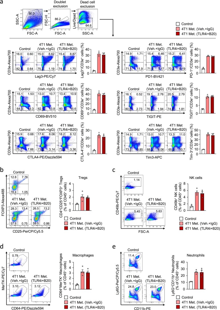
Extended Data Fig. 9. Immune response in metastatic lungs treated with inhibitors of TLR4 and VEGF.
a, Flow cytometry analysis of T cell exhaustion in control lungs or lungs from 4T1 metastasis-bearing BALB/c mice after treatment with TLR4i and anti-VEGF (B20). Following exhaustion markers were analyzed on T cells: Lag3, CD69, CTLA-4, PD-1, TIGIT and Tim-3. Shown are flow cytometry examples and quantification of the indicated exhaustion markers expressed by T cells. Data are means with s.e.m. b-e, Flow cytometry analysis of regulatory T cells (Tregs, CD25+/FOXP3+), natural killer cells (NK cells, CD49b+), macrophages (CD64+/MerTK+) and neutrophils (Ly6G+/CD11b+) in control lungs or lungs with 4T1 metastasis in BALB/c mice upon treatment with TLR4i and B20. Shown are flow cytometry examples (left) and quantifications as means with s.e.m. (right). Analysis was performed on CD4+ gated cells (Tregs analysis) or CD45+ gated cells (analysis of NK cells, macrophages or neutrophils). Control group, n= 3 mice and metastasis groups treated with vehicle/IgG or TLR4i/B20, n= 4 mice each group.

