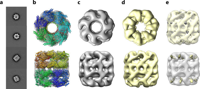
Fig. 2. 3D reconstruction of GroEL complexes that were either matrix-landed from the ion beam of a modified Orbitrap mass spectrometer or conventionally prepared.
a 2D Class averages obtained from negatively stained matrix-landed GroEL cations. b Top and bottom views of a three-dimensional reconstruction of GroEL made from the particles contained within the class averages shown in a, fit to a previously determined GroEL structure (PDB:5W0S). c Top and bottom views of (b) without ribbon model. d Top and bottom views of conventionally prepared and imaged GroEL. e Difference maps of matrix-landed and conventionally prepared GroEL. Top structure in (e) presents the subtraction of the conventionally prepared model from the landed model (where gray indicates difference) while the bottom displays the reverse (yellow indicating difference).

