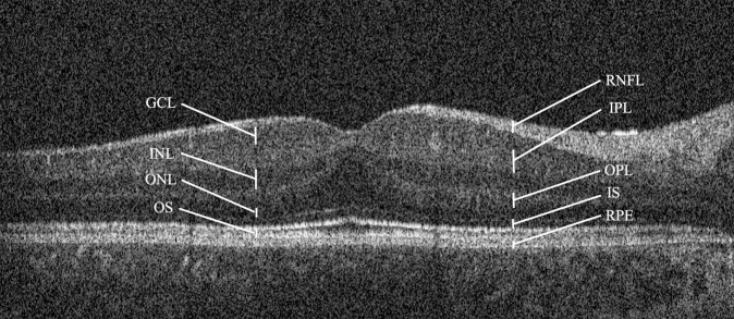
Fig. 1. Flattened horizontal OCT scan through the fovea of a 5-year-old subject with Down syndrome.
Grade 1 foveal hypoplasia is demonstrated, with continuation of the inner retinal layers through the fovea. The retinal layers are labelled as follows: retinal nerve fibre layer (RNFL), ganglion cell layer (GCL), inner plexiform layer (IPL), inner nuclear layer (INL), outer plexiform layer (OPL), outer nuclear layer (ONL), inner photoreceptor segments (IS), outer photoreceptor segments (OS), and retinal pigment epithelium (RPE).

