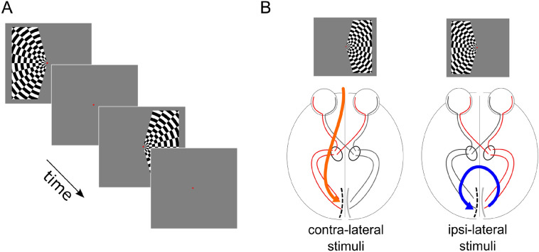
Fig. 1.
Visual paradigm. Panel A: left or right visual hemifields were stimulated by a contrast-defined lateralized dart-board pattern, alternated with periods of gray screen, which constituted the baseline condition (Tootell et al. 1998; Gouws et al. 2014; Fracasso et al. 2018). Panel B: participants were asked to fixate on the center of the screen and maintain stable fixation on the red center dot. The alternate segments of the pattern moved in opposite radial directions and the motion direction changed unpredictably. In participant 1 (P1) the electrode grid was placed on the left hemisphere (see dashed line in the sketched primary visual cortex in panel B); in participant 2 (P2) the grid was placed over the right hemisphere (not shown). Thus, the ipsi-lateral and contra-lateral conditions were associated with opposite hemispheres in the two participants. The ipsi-lateral and contra-lateral conditions were defined with respect to the placement of the ECoG grid. In the example above (P1, ECoG grid in the left occipital pole) the contra-lateral stimuli response is located in the right hemifield, the ipsilateral stimuli response in the left hemifield. The opposite mapping occurred for P2, where the ECoG grid was placed on the right occipital pole

