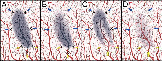
Fig. 3.
Illustration of different perfusion patterns with or without anterograde collaterals. (A) The retrograde (blue arrows) and anterograde (yellow arrows) were markedly decreased and a hypoperfusion pattern was observed. (B) The anterograde collaterals were significant reduced and perfusion depended on the retrograde collaterals. This was observed in both hypoperfusion and compensated perfusion patterns. (C) The anterograde collateral was prominent and this pattern was observed more often in compensated perfusion and less often in hypoperfusion patterns. (D) Both anterograde and retrograde collaterals were present, representing a normal perfusion pattern. This illustration was
modified from the figure by Rudilosso et al. [14]

