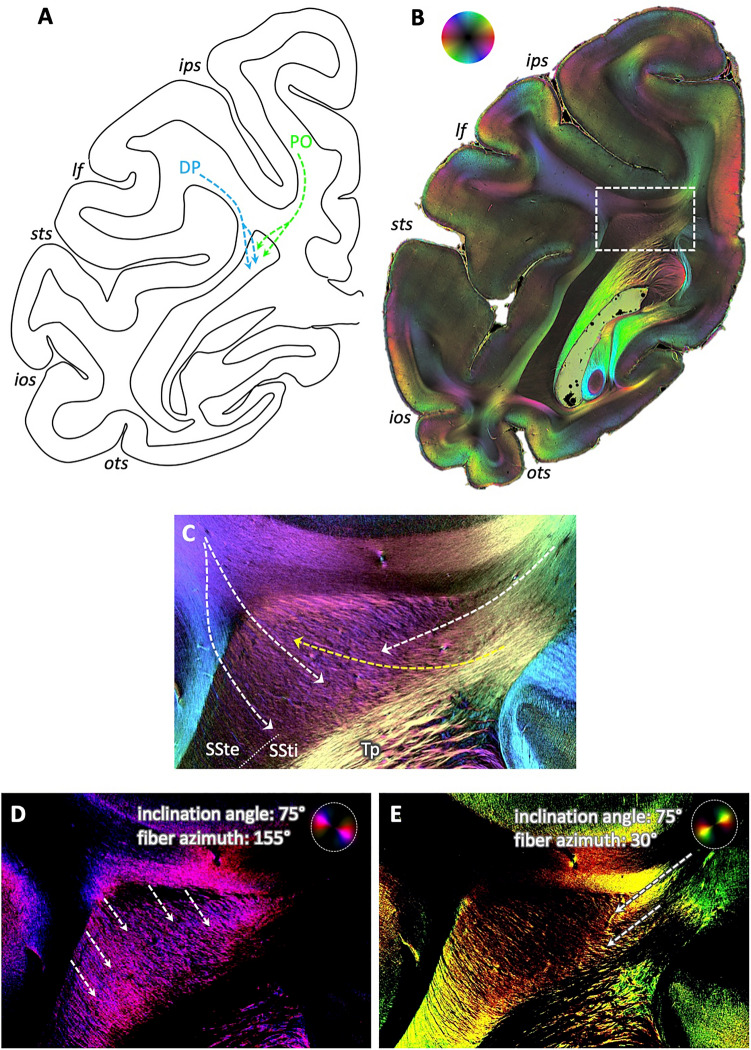
Fig. 5.
Separate entrance of fibers within the dorsal sagittal stratum of the vervet monkey brain. A Schematic depiction of routes of tracers after injection in areas DP (blue arrows) and PO (green arrows), according to Schmahmann and Pandya 2006 (their cases 18 and 17, respectively, p. 245). Note the separate entrance of the fiber bundles into the sagittal stratum. B 3D-PLI-derived FOM at a comparable coronal sectioning plane as in A. Region of interest in dorsal sagittal stratum as analyzed in C indicated by a white box. Color sphere indicates color coding of fiber orientations. C Enlarged, contrast-enhanced depiction of fiber architecture within dorsal sagittal stratum. White arrows indicate the predominant courses of the fibers, from top left into the sagittal stratum, reflecting connections from dorsal lateral parieto-occipital cortex (area DP), and from top right, reflecting connections from mesial parietal cortex (area PO). Note the crossing of the transcallosal fibers from the tapetum through the sagittal stratum to the cortex (yellow arrow). D and E Same enlarged view as in C, but as flashlight views of particular fibers of given direction within the section and inclination as indicated by the color coding within the panels. lf lateral fissure. For other conventions, see Fig. 2

