Fig. 23.

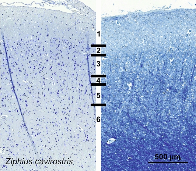
Photomicrograph of Nissl (left) and Kluver–Barrera (right) stain of the primary visual cortex of Cuvier’s beaked whale (Ziphius cavirostris). The images have been voluntarily slightly overexposed to enhance the structure visualization

Photomicrograph of Nissl (left) and Kluver–Barrera (right) stain of the primary visual cortex of Cuvier’s beaked whale (Ziphius cavirostris). The images have been voluntarily slightly overexposed to enhance the structure visualization