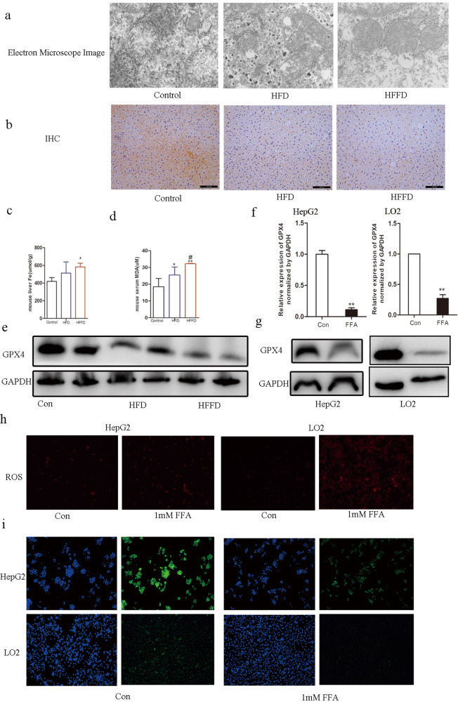
Fig. 3. Ferroptosis in mice with NAFLD and FFA-induced hepatocytes.
a Electron microscope image of the liver in mice with HFD, HFFD, or a regular chow diet. b IHC analysis of GPX4 expression of the liver in mice with HFD, HFFD, or regular chow diet. c Liver Fe in mice with HFD, HFFD, or regular chow diet. d Serum MDA in mice with HFD, HFFD, or regular chow diet. e Western blot analysis of GPX4 levels in mice with HFD, HFFD, or regular chow diet. f Relative mRNA expression levels of GPX4 in FFA-induced HepG2 and LO2 cells, **compared to control, p < 0.001. g Western blot analysis of GPX4 levels in FFA-induced HepG2 and LO2 cells. h ROS expression in FFA-induced HepG2 and LO2 cells. i IF of GPX4 expression in FFA-induced HepG2 and LO2 cells.

