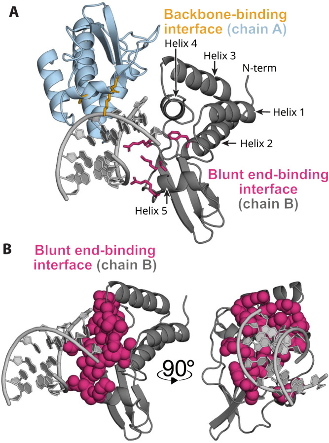
Fig. 1. VP35 dsRNA interactions occur primarily through flat interfaces.

A Crystal structure of two copies of VP35’s IID (dark gray and light blue) bound to dsRNA (light gray) via two flat interfaces (PDB ID 3L25). Mutations to residues highlighted in pink and yellow sticks eliminate dsRNA binding. B Isolated chain B from the same view as panel A and after 90° rotation in the Y axis now highlighting the dsRNA interacting VP35 surface in the blunt-end-binding protomer. The blunt-end-binding interface (pink, 3L25 chain B) is shown as spheres to highlight that VP35 lacks deep pockets amenable to binding small molecules.
