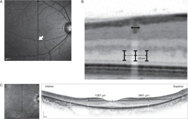
Fig. 2. Measurement of ONL thickness with SD-OCT imaging.
Measurement and analysis of outer nuclear layer (ONL) thickness with the vertical line scan spectral-domain optical coherence tomography (SD-OCT) imaging. A SD-OCT 30 degrees vertical line scan imaging through the fovea was examined. B 50–100 μm retinal vessels were selected. We manually measured ONL thickness under the vessel. Note that unsmoothed images were used to determine the border of the layer precisely. As a control, we measured ONL thickness in adjacent areas: 100 μm superior and inferior from the centre of the vessel. C In patients with EYS-associated retinitis pigmentosa (RP), superior and inferior ellipsoid zone (EZ) length from the fovea was manually measured with the spectral-domain optical coherence tomography (SD-OCT) image.

