
Fig. 1.
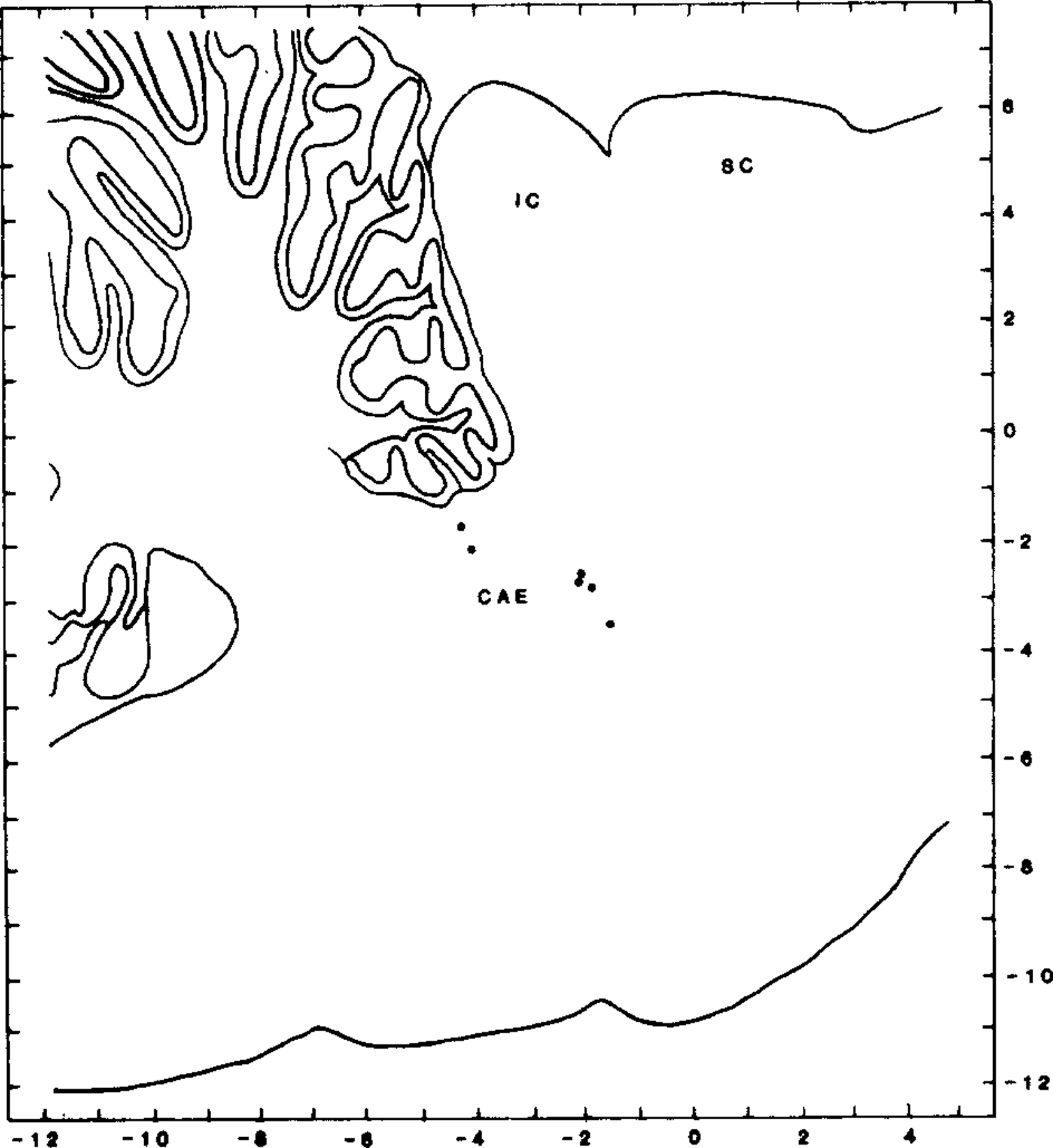
Distribution of recorded cells within the dorsolateral pons. Cells are plotted on sagittal section, 2.9 mm lateral to midline. Laterality of recorded cells ranged between 2 and 3.5 mm from midline. CAE indicates position of locus coeruleus; IC, inferior colliculus; SC, superior colliculus.