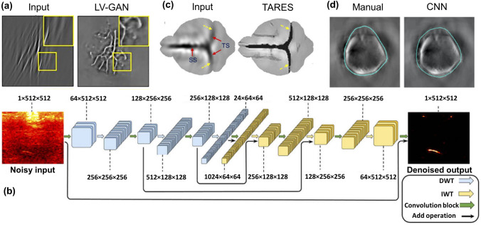
Fig. 3.
a Reconstructed limited view vessel phantom image using LV-GAN. Reprinted with permission from Wiley–VCH [101]. b Schematic of the MWCNN architecture employed to improve contrast in low fluence PAT settings. U-Net employed for bandwidth enhancement in PAT. Reprinted with permission from OSA [110]. c Tangential resolution improved in vivo PAT brain image using TARES network. Reprinted with permission from OSA [117]. d Boundary segmented in vivo mouse liver images using CNN. Reprinted with permission from SPIE [132]

