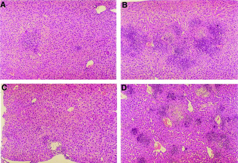
FIG. 2.
Histological analysis of the livers of L. monocytogenes-infected mice (3.7 × 10⁴ CFU/animal). Shown are representative liver sections fixed in 2% paraformaldehyde buffer solution, embedded in paraffin, and stained with hematoxylin and eosin from anti-IL-10R-treated mice on days 3 and 5 (A and C, respectively) postinfection and the respective controls on days 3 and 4 (B and D, respectively).

