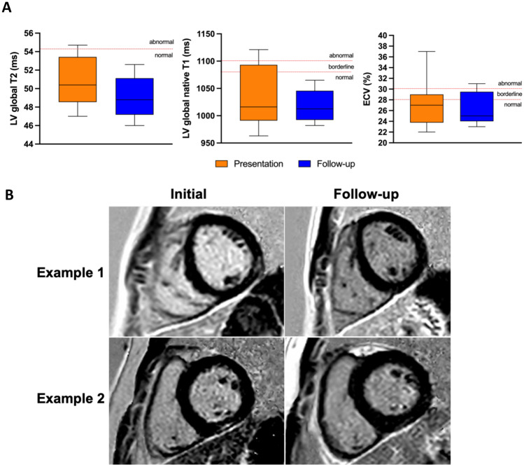
Fig. 1.
A Left ventricular (LV) global T1, LV global T2, and extracellular volume (ECV) on CMR at presentation and follow-up in children with vaccine-associated myocarditis. B Cardiac magnetic resonance short-axis views at presentation and follow-up. Example 1: LGE in the mid-wall of the mid left ventricle at the junction of the anterolateral and inferolateral segments, which completely resolved on follow-up. Example 2: late gadolinium enhancement (LGE) in the inferior basal segment, with significant improvement but slight persistent findings on follow-up

