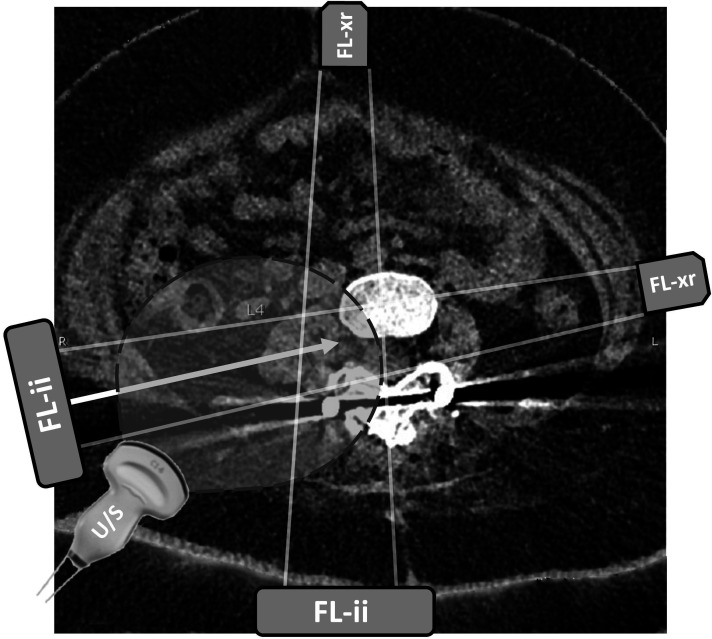
Figure 1.
Axial CT view at the level of lumbar vertebra 4. Axial CT image shows locations of the lumbar spine, with planned needle trajectory shown with the solid arrow. Projected needle path crosses the posterior aspect of the peritoneum but avoids the bowel and other viscera. Atrophic psoas muscle is seen immediately lateral to the spine. Fluoroscopy beam positions for transforaminal lumbar puncture are shown with source (FL-xr) and image intensifier (FL-ii) in tunnel view and anterior–posterior view. Curvilinear ultrasound (U/S) probe position and field of view are drawn over the image. Image included with patient assent and parental consent. LP, lumbar puncture.

