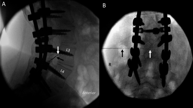
Figure 2.
Needle advancement using fluoroscopic guidance. A thin spinal needle enters the spinal canal via transforaminal approach through an introducer needle in lateral tunnel (A) and anterior–posterior (B) views. Arrows show positions of the introducer needle tip (black) and the thin spinal needle tip (white). Image included with patient assent and parental consent. R, right.

