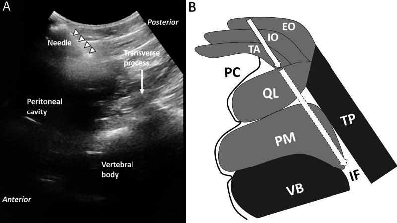
Figure 3.
Ultrasound view. (A) Ultrasound view of needle trajectory. Curvilinear robe is oriented in an axial in-plane view to observe the introducer needle and its anticipated trajectory toward the spine. In this example, the needle trajectory extends just along the posterior border of the peritoneum toward the foramen. (B) Drawing of structures shown in A. Solid border on arrow shows the needle advanced to the depth shown in A. Dashed border on arrow shows the anticipated trajectory of the needle into the foramen. Image included with patient consent. EO, external oblique; IF, intervertebral foramen; IO, internal oblique; PC, peritoneal cavity; PM, psoas major; QL, quadratus lumborum; TA, transversus abdominis; TP, transverse process; VB, vertebral body.

