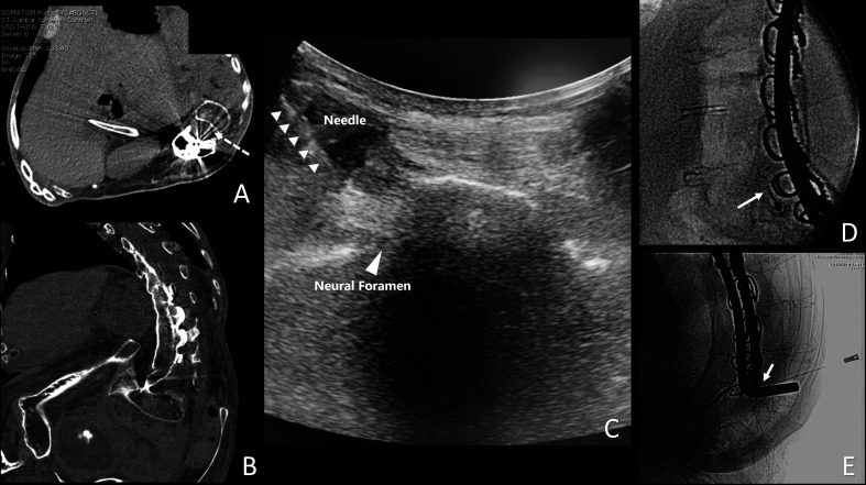
Figure 4.
Multiple views of the spine in a patient undergoing transforaminal spinal anesthesia. (A) Planned transforaminal needle trajectory (dashed arrow) from an axial view from a prior CT scan. (B) Far left: lateral position of the spine in a coronal view from a prior CT scan. (C) Ultrasound view of the needle trajectory. (D) Lateral tunnel fluoroscopic view. (E) Anterior–posterior fluoroscopic view of a 25-gage 5 cm non-cutting needle used for spinal anesthesia. Image included with patient consent.

