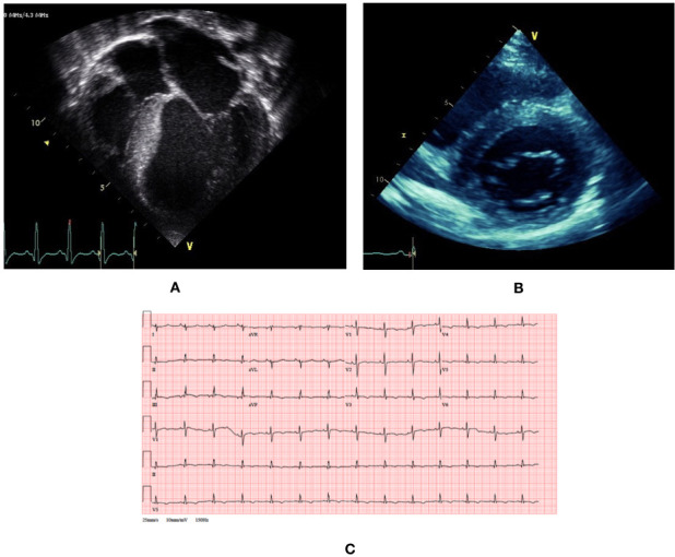
Figure 2.

Clinical phenotype of a patient with atypical presentation of Friedreich’s ataxia-associated hypertrophic cardiomyopathy. (A) Transthoracic echocardiogram at presentation at age 5 shows dilated and hypertrophied phenotype with impaired systolic function. (B) Transthoracic echocardiogram at age 15 shows concentric hypertrophy with maximal left ventricular wall thickness of 14 mm. (C) 12-lead ECG at age 15 shows small voltages, right axis deviation and widespread repolarisation abnormalities (flat or inverted T waves inferiorly and V2–V6).
