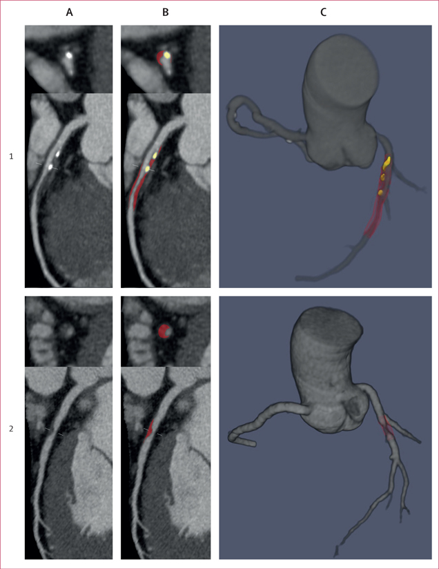
Figure 2. Case examples of deep learning plaque segmentation.
(A) Curved multiplanar reformation coronary CT angiography images showing lesions in the proximal-to-mid left anterior descending artery (1) and the mid left anterior descending artery (2). (B) Deep learning segmentation of calcified plaque (yellow) and noncalcified plaque (red). (C) Three-dimensional rendered view of the coronary tree showing deep learning plaque segmentation in the individual analysed segments. All lesions in each vessel were analysed by deep learning and measurements summed on a per-patient level.

