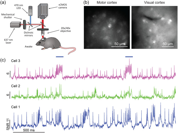
Fig. 11.
Single cell voltage imaging in awake mice using a wide-field imaging setup. (a) Experimental setup: awake mice were head-fixed under a wide-field microscope. (b) Representative SomArchon-expressing neurons visualized via EGFP fluorescence in cerebral cortex. (c) Voltage imaging time-courses from three representative neurons in hippocampus; horizontal bars denote optogenetic stimulation. Reproduced with permission from Piatkevich et al.99

